— Research Projects
Microtubules in Brain Development and Disease.

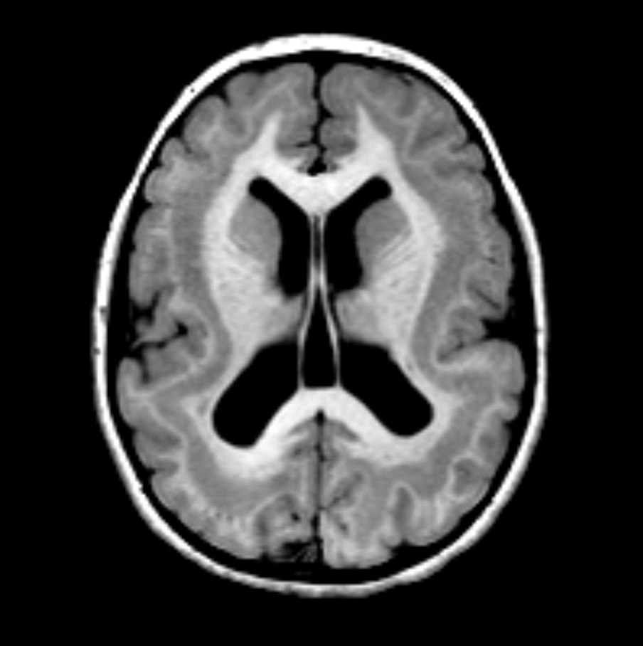
The life of a neuron involves many movements and morphological changes. Newborn neurons often migrate long distances to reach their final location in the developing brain. The neuron extends neurites, the axon generates a synapse, and the other neurites extend to form the complete neuritic arbor, which is pruned and reformed during development and learning. Brain development and function depend on the ability of neurons to accomplish these rearrangements. Each of these morphological changes require the active remodeling of the neuron's microtubule cytoskeleton. Each task is performed by microtubule associated proteins (MAPs). Misregulation or failure of MAPs causes a spectrum of disorders in brain development and neurodegenerative disease. Our lab studies the MAPs that cause these diseases. What do these MAPs do to microtubules and why do mutations cause disease? A current focus in the lab is the MAP doublecortin (DCX). Mutations in DCX cause an inherited form of epilepsy known as double cortex syndrome. Effective diagnosis, treatment planning, and drug development for these diseases requires a precise understanding of the molecular mechanisms underlying microtubule remodeling and MAP function in neurons.
Biophysics of Microtubule Polymerization
At the most fundamental level, how do tubulin dimers assemble into a properly-formed microtubule? Our lab investigates the biophysical basis of tubulin polymerization by measuring the kinetics and energetics of microtubule formation. What is the threshold that gates the addition of a new tubulin dimer to the end of a microtubule and how do MAPs modulate these thresholds? Our experiments provide us with a nano-scale view of polymer formation and, importantly, allow us to understand MAP function at the nano-scale as well. We obtain a deep understanding of MAP function and the specific breakdown that arise from disease-causing mutations.
Why single molecules and molecular biophysics?
In experiments with a large number of microtubules, such as observations of a dense cytoskeleton in cells or bulk biochemical assays, several distinct molecular mechanisms can produce identical results. For these reasons, single molecule experiments have taken hold of the cytoskeleton field. Single molecule experiments have deep roots in the study of microtubules. A short list of the discoveries made by observing single microtubules in vitro includes: microtubules undergo "dynamic instability'', kinesin walks processively on microtubules, and microtubule severing enzymes cut microtubules in the middle. Looking at recent examples, single molecule experiments have defined the hand-over-hand model for kinesin motility, demonstrated that depolymerizing enzymes remove tubulin from microtubule ends, and that proteins such as EB1 can autonomously track microtubule plus-tips. The reconstitution of microtubule growth and shrinkage in vitro has been a rewarding development. Application of these techniques to proteins involved in brain development and disease is the most effective way to advance our understanding of these regulators of neuronal morphology.